
Indietro
Ultimo aggiornamento:
5 display hurdles in mammography - and the solution
Settore sanitario · Tumore al seno · 4 minuti di lettura
Breast imaging is a specialty with high requirements. Every detail is delicate, so the way you view your images is crucial. This results in the fact that it's not always easy to find a solution that works best for you, as there are many things to take into account. Based on our deep-rooted knowledge of the field, we've listed some of the main hurdles breast radiologists experience.
Hurdle 1: Suboptimal reading conditions
The field of breast imaging is often described as a challenging and disconnected environment, especially since radiologists are required to view more and different types of imaging modalities. Different sets of images have their own specific display specifications, compelling radiologists to use multiple workstations. This has an impact on radiologists’ efficiency and productivity.

Hurdle 2: Lack of color
Radiologists are also reviewing images from supporting modalities – such as PET, US, MR or NucMed – that require monitors with color display. Breast US and breast MR, for example, are taken to further refine the diagnosis. Being able to view Color Doppler US, breast MR and standard mammography images simultaneously on one display supports radiologists in accurate decision-making and improving health outcomes.
Hurdle 3: Limited screen space
Breast imaging radiologists are often limited in the real estate available on the monitors, which restricts their view of full complete images. Today, radiologists need to pan, zoom, and rearrange images in order to get the best image for analysis. It’s no wonder that having access to life-size images can impact diagnostic interpretation.

Our Coronis OneLook monitor allows images to be viewed in full resolution, no panning or zooming needed.
Hurdle 4: Challenges of digital breast tomosynthesis
Digital breast tomosynthesis can take a toll on radiologists as the program requires scrolling through 50-100 slices of breast tissue for each of the four views of a normal screening mammogram. Even though studies have shown breast tomosynthesis increases cancer detection and reduces unnecessary callbacks, these results do not come without impacting radiologists’ workflow.
Hurdle 5 – Lack of workflow tools
Tasked with reading dozens of studies per day, radiologists are in desperate need for tools that automate routine tasks and help ensure a more productive, time-efficient way of working. Additionally, radiologists are prone to sitting in a dark environment to view images, affecting their reading comfort. Tools that support better viewing ergonomics (e.g. adjustable ambient lighting) are no unnecessary luxury!
Sembra che tu non abbia accettato i cookie
Attiva i cookie di performance per guardare questo video.
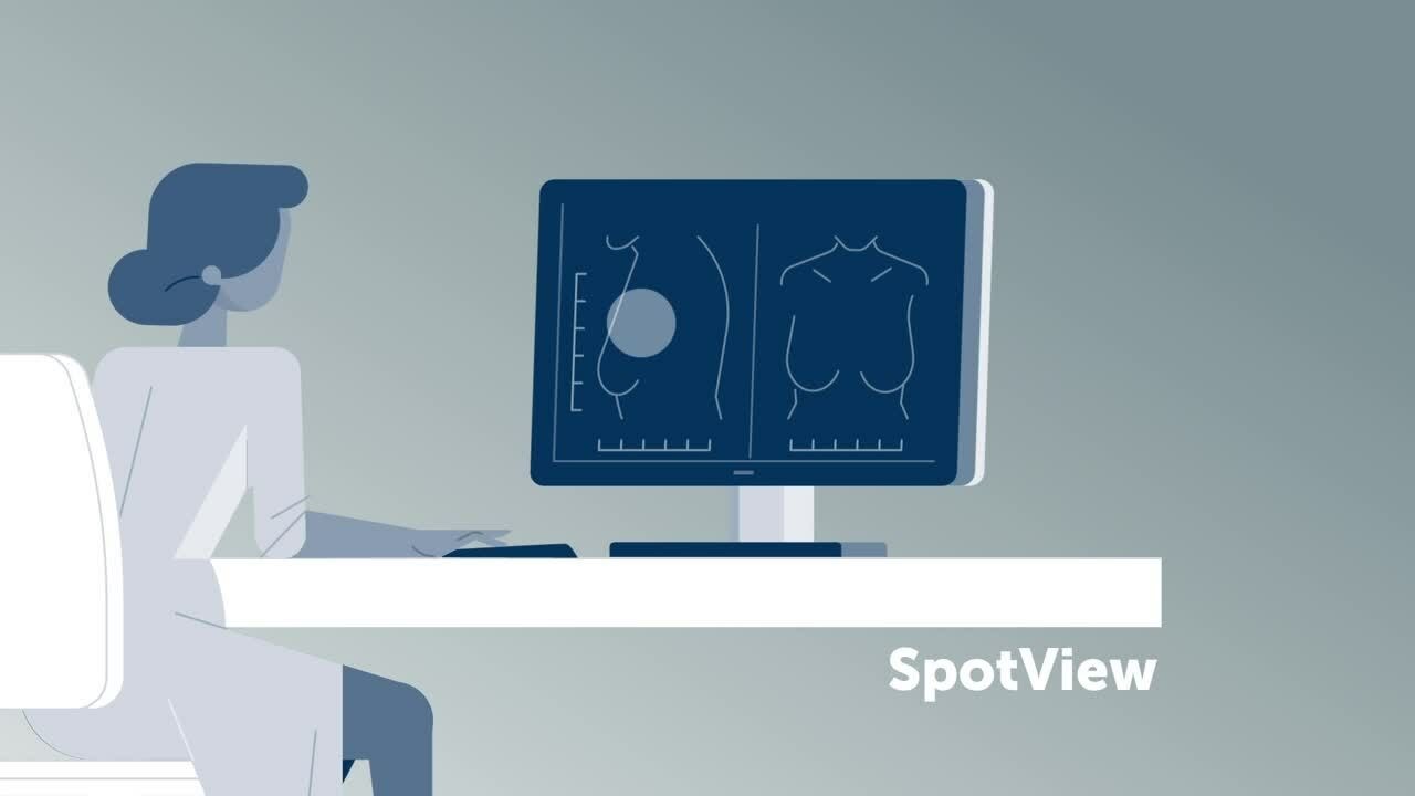
Our SpotView technology focuses the light on an area of interest.
Discover our display solutions for breast radiology
There are many obstacles in breast imaging, but our breast imaging solutions are grounded in a long history of research. With dedicated monitors for traditional digital mammography, multi-modality breast imaging, and breast tomosynthesis, we aim to set the standard of care for women's health.
- We offer multi-head displays, in addition to a range of Fusion displays that allow to portray multiple images at once.
- Our medical display controllers make it possible to use our Intuitive Workflow Tools, designed to optimize your reading experience and working comfort.
- Our RapidFrame technology enables you to scroll or cine through large sets of tomosynthesis slices without lag or blurring of the image.
Related articles


Precedente
di
...
Successivo